Biosecurity and Bionanosciences Group
Our group conducts bionanocience research that applies nanoscience and nanotechnology to problems for national biosecurity interests. We are a multidisciplinary team with expertise in physics, chemistry, materials science and biology. This unique cross cutting expertise allows us to work together on basic and applied research toward LLNLʼs mission in nonproliferation, counterterrorism and life sciences. Our current research focus includes developing novel detection methods for biological agents, advanced bioanalytical and molecular imaging instrumentations for nanoscale characterization, novel carbon nanotube fabrics that repels chemical and biological agents and nanolipidprotein technology as a medical countermeasure to biological threats.
Research Areas
-
Chemical threat responsive carbon nanotube (CNT) membranes
-
Nanolipoprotein particles (NLPs) as an in vivo delivery platform for biomedical applications
-
Biomineralization and crystal growth
-
Chemical mapping by imaging mass spectrometry
-
Tracking aerosol airflow patterns - DNATrax
-
Point-of-care diagnostics for infectious disease detection
-
Study biological response in cells

Dynamic Multifunctional Materials for a Second Skin
Contact: Francesco Fornasiero
Chemical threat responsive CNT membranes
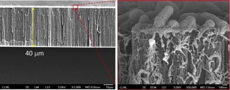
Several military and civilian settings require the use of protective clothing to shield the individuals from chemical agents, dusts, organic and biological hazards. Wearing conventional protective equipments in these conditions restricts body evaporative heat loss with the risk of heat exhaustion and heat stroke. To address this problem, we are developing highly-breathable responsive membranes having a-few-nm wide vertically-aligned carbon nanotubes as only through pores. Because of the small pore sizes, these membranes provide an effective barrier against biological threats. To protect from chemical warfare agents (CWA), we are developing CWA-responsive surface-coatings that sense and block the transport of CWAs through the pores upon threat exposure, or exfoliate upon reaction with CWAs.
see LLNL News Releases: New military apparel repels chemical and biological agents
Nanolipoprotein particles (NLPs) as an in vivo delivery platform for biomedical applications
Contact: Craig Blanchette
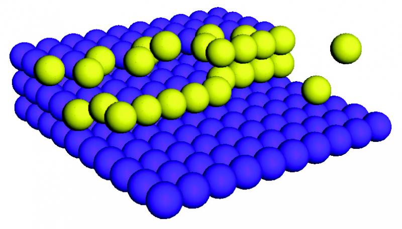
Development of nanolipoprotein particles (NLPs), also known as nanodiscs and reconstituted High Density Lipoproteins (rHDLs), for in vivo delivery applications. NLPs are biocompatible particles analogous to high-density lipoproteins (HDLs). They are nano-scale (10-25 nm) discoidal membrane bilayer mimetics that form through spontaneous self-assembly of purified lipoproteins and lipids. The versatility of the NLP self-assembly reaction allows for the incorporation of lipids bearing functional groups at the solvent interface of the lipid bilayer, enabling multifunctional NLPs to be prepared in a single step. Thus, myriad functional groups can be incorporated into the NLP, including chemical groups enabling covalent and noncovalent conjugation of biomolecules.
We have previously focused primarily on utilizing NLPs for vaccine applications and exploited the multifunctional nature of the NLP to develop a variety of methods to incorporate adjuvants and antigens on a single particle, which has allowed us to achieve co-localized delivery of these two components. In animal experiments, we have shown the co-localized delivery of adjuvants and antigens significantly enhances both the innate and adaptive immune response. Our efforts have focused primarily on relevant pathogens. During the course of this work we have also developed strategies to engineer the NLP platform to modulate the immune system, which can be used as a standalone therapeutic. More recently we have initiated studies to evaluate the potential of using this platform for targeted and tissue specific delivery of therapeutics with an emphasis on bladder cancer. Through this work we have been able to successful engineer the NLP to be stable for up to 24 hrs in blood serum, while incorporating both a targeting ligand and therapeutic payload. Although nanoparticles in general have been evaluated as in vivo delivery vehicles over the past few decades, this particular platform is relatively new to this field and holds great promise for a wide range of biomedical applications.

Understanding the physical mechanisms of biomineralization at the nanoscale
Contact: Chris Orme
Studies the fundamental physics of crystallization and materials assembly with application to biomineralization, biomimetic synthesis, controlled synthesis of nanostructures, catalysis, interfacial dynamics, and electrochemical deposition.
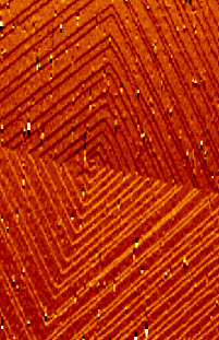
The atomic force microscope (AFM) has been used since 1986 to produce topographic maps of nanostructures. An ultrasharp tip scans across a sample’s surface, and a computer records the path of the tip, slowly building a three-dimensional image. When force and displacement data are combined in various algorithms, the resulting calculations reveal information on mechanical properties, such as hardness, and any other reactions to an applied force.
Unlike the scanning electron microscope, which provides a two-dimensional image of a sample, the AFM provides a true three dimensional surface profile. Additionally, samples viewed by AFM do not require special treatment that would destroy the sample. AFMs also do not need to operate in a vacuum environment, as do electron microscopes. The AFM can be used on materials that don’t conduct electricity. The microscope is unique in its ability to measure the mechanical properties of both hard and soft materials. The latter has traditionally been more difficult because soft materials, such as biological tissues, are part solid and part fluid.
Although the AFM produces a smaller image area than electron microscopes (micrometers versus millimeters, respectively), it can image and manipulate matter at the nanoscale, allowing researchers to both study a molecule’s structure and properties and organize molecules into predetermined patterns.
Molecular Imaging of Biological Systems by Mass Spectrometry
Contact: Kuang Jen J. Wu
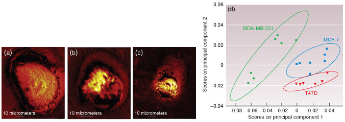
We are developing integrated imaging mass spectrometry (IMS) technology to perform chemical imaging of analytes on cells and tissue surfaces. Direct tissue profiling by IMS provides detailed mapping of the complex molecular pattern across a tissue sample. The methodology generates a collection of mass spectra from a defined sample surface. Instead of interpreting these information-rich spectral set visually, we have employed multivariate statistical analysis tool to analyze the entire collection of chemical maps. Our approach combines high spatial resolution with non-supervised spectral data processing technique. Based on the spectral patterns the correlation between biologically relevant molecular compositions and their spatial distribution can be visualized directly and intuitively. The combined toolbox allows us to perform hyperspectral visualization of complex IMS data set obtained from biological samples. Profiling and IMS have been used to characterize multiple biological systems. We have successfully demonstrated differentiation of cancer cell types and separation of benign from malignant biopsy tissues.
DNATrax
 Contact: George Farquar
One of the most overlooked threats to human health is poor indoor air quality. Various air pollutants exist indoors, including biological pollutants (molds, bacteria, viruses, pollen, animal dander, dust mites, etc.), second hand smoke, combustion pollutants, and other chemicals (formaldehyde, asbestos, radon, etc.). With inadequate ventilation, these contaminants become concentrated and can result in detrimental effects on human health. Pinpointing areas where pollutants have accumulated requires an understanding of the airflow in a given space.
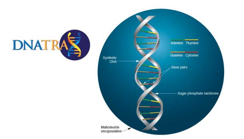
DNATrax, a 2013 R&D 100 Award winning technology, is a safe and versatile material containing food-based microparticles that can be used for the safe and effective detection and tracking of aerosol releases in both interior and exterior environments. By combining FDA-approved sugars, and a unique non-biological DNA bar code, a microparticle that simulates the aerosols compromising the air around us was produced. DNATrax will enable pollutant identification and empower customers to improve indoor air quality.
see LLNL S&TR article: DNA tagged sugar particles simulate aerosol ariflow patterns
Point-of-Care Diagnostics for Infectious Disease Detection
Contact: Brian Baker
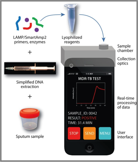
(A) Mobile Phone Platform for Genomic Disease Detection
Although tuberculosis (TB) is the second deadliest infectious disease worldwide, there are no TB diagnostics that combine speed, accuracy and low cost. In collaboration with UC Berkeley and UC San Francisco, we are developing a portable, point-of-care device for diagnosis of TB and associated drug resistance.
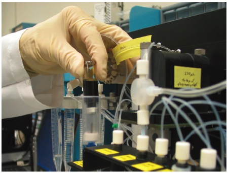
(B) Point-of-Care Medical Device for Diagnosis of Sepsis
For the septic patient, rapid identification of the causative agent and administration of the proper antibiotic is critical to patient outcome. We developed a portable, automated system for the purification, amplification and detection of bacterial DNA from human whole blood, via Loop-Mediated Isothermal Amplification.
Utilizing biophysical methods to study biological response in cells
Contact: Craig Blanchette
We have developed biophysical methods to measure mechanical and structural changes in cells under physiological relevant conditions. These methods primarily utilize combined atomic force microscopy (AFM) and confocal fluorescent microscopy (CFM). This work has focused primarily on two projects: 1) measuring changes to cytoskeletal elements in response to localized delivery of pathogenic stimuli (Internalin A (InlA) from Listeria monocytogenes) and 2) measuring the mechanical stiffness while imaging changes to the cytoskeletal elements of chondrocytes in the presence and absence of cytoskeletal disruptors.
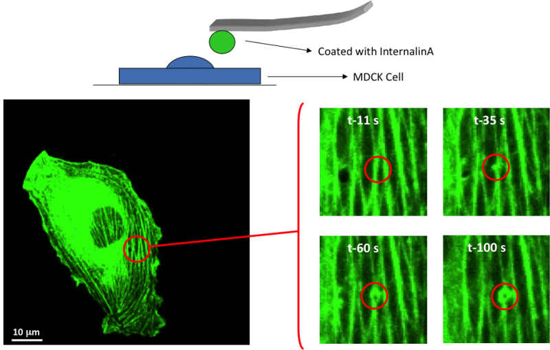
Video: Probing and individual cell with a pathogenic mimetic and measure the cytoskeletal response.